How we portray and engage with the world around us is fundamentally influenced by our sense of vision. The visual area is located in about one-third of the cortical surface of the primates. Vision loss, either at birth or much later in life, affects how we perceive the world. It is intriguing to know how the brain of sighted and blind individuals differ. In the paper "The nature of consciousness in the visually deprived brain," the authors Ron Kupers, Pietro Pietrini, Emiliano Ricciardi, and Maurice Ptito discuss various topics pertaining to the nature of consciousness in those without vision. This research answered an important question:
What can we learn about the functional development of the human brain in physiological conditions by studying blindness?
(Source: https://doi.org/10.3389/fpsyg.2011.00019)
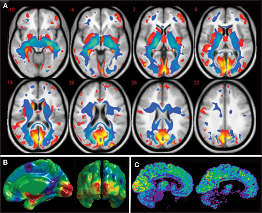
Apart from this, recent years have seen the successful application of brain imaging methods based on magnetic resonance imaging (MRI) for the investigation of changes in grey and white matter in the brain of a blind subject. Some of the results can be observed in the figure below for example:
- (A) Congenitally blind people have less grey (red), and white (blue) matter in their axial brain slices than matched sighted control participants. The volume of every element of the blind person's visual system is diminished (Ptito et al., 2008b).
- (B) Cortical thickness variations between sighted control participants and congenitally blind individuals. Congenitally blind patients had thicker cuneus cortex despite having a smaller occipital cortex in size.
- (C) Congenitally blind brains have a higher resting-state glucose metabolism, as shown by mid-sagittal brain slices.
 |
| (Source: https://doi.org/10.3389/fpsyg.2011.00019) |
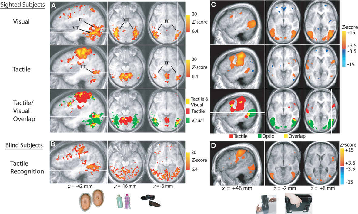
Section (A, B) in the above figure shows the extrastriate ventral temporal cortex's supramodal neuronal response. Examples of stimuli utilized during tactile and visual recognition of several object categories by sighted and congenitally blind subjects include life-size masks of faces, plastic bottles, and shoes. It is observed in the above figure in sections (A, B) that tactile and visual object perception activates the inferior temporal (IT) and ventral temporal (VT) areas. The tactile/visual overlap map displays the regions that are triggered by both tactile and visual perception (represented in yellow), as well as the regions that are only activated by touch and visual perception (shown in red and green, respectively).
Section (C, D) in the above figure shows the supramodal neuronal response in the hMT+ cortex. The hands of the subjects were placed on braille-like dot patterns. In the figure, the brain regions that responded during the perception of tactile or optic flow in sighted participants and during the perception of tactile flow in blind subjects are displayed. Areas that are activated by both tactile and optic flow perception are represented in yellow on the tactile/visual overlap map. In contrast, areas that are only activated by tactile and optic perception are depicted in red and green, respectively.
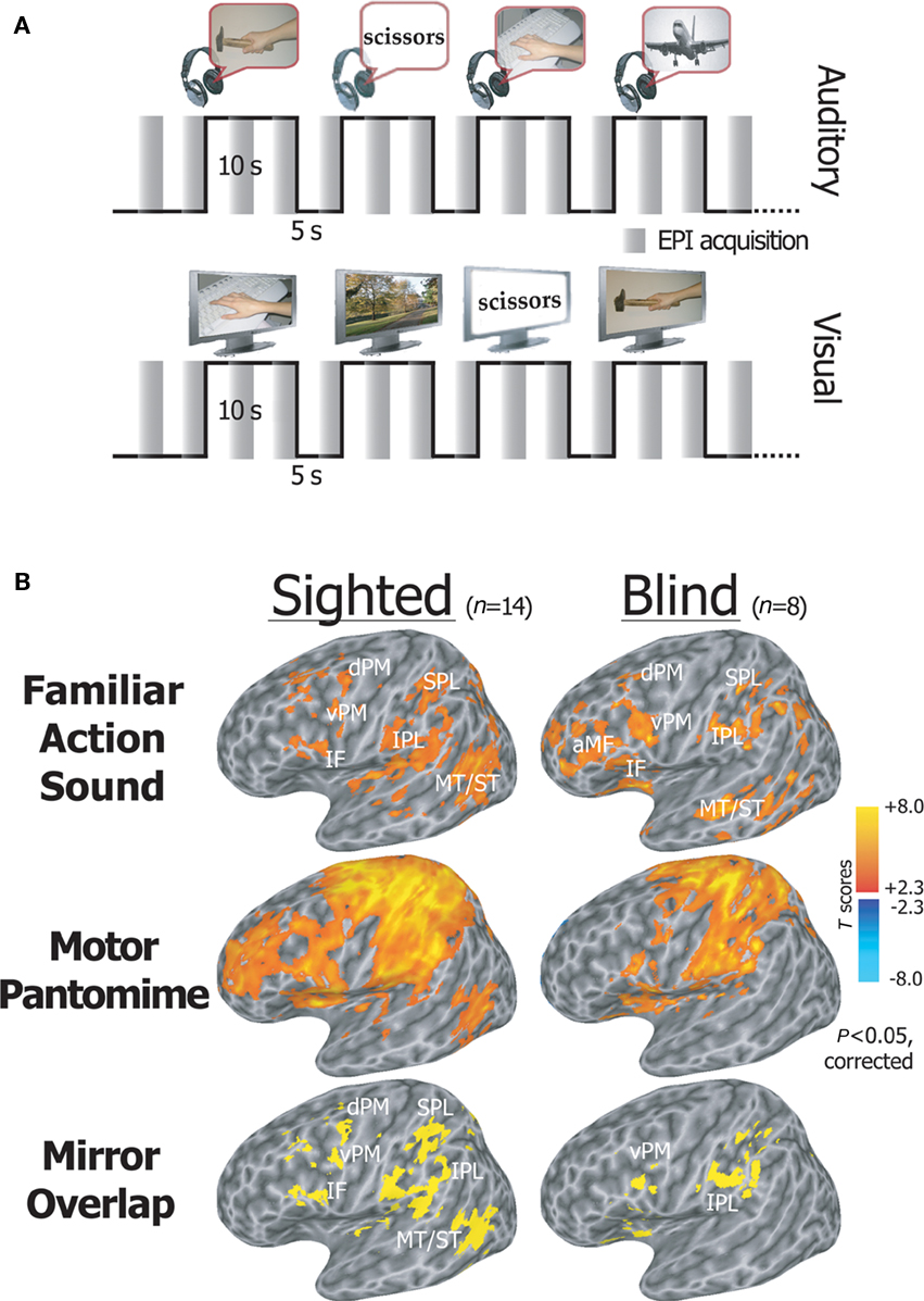
Also, using fMRI, a similar "mirror" system has been found in humans. It is believed to be important for language development, learning by imitation, and comprehending behavior and intention. The inferior frontal cortex, together with the occipital, temporal, and parietal areas, are part of a complex network that is activated by the human mirror neuron system when it detects actions performed by others. A plethora of research has been conducted in order to fully comprehend the functioning of the mirror system; however, do not address the issue of whether the activation of the mirror system in response to acoustically presented stimuli may be mediated by the acoustically evoked visual imagery of the depicted action or is rather independent of vision. To this extent, congenitally blind and sighted volunteers underwent a series of tasks that alternated between the sporadic display of manually performed action or background sounds/movies and a motor pantomime of a "virtual" tool or object manipulation task in order to measure neural activity using an fMRI sparse sampling block design as shown in the figure below (A). The brain's activity is seen on statistical maps (B) during familiar action listening compared to environmental noises, and during the action, motor pantomime compared to rest.
 |
| (Source: https://doi.org/10.3389/fpsyg.2011.00019) |
We live in a world where vision plays a major role. The visual cortex in primates makes up around 30% of the overall cortical surface. As a result, losing one's vision is among the most disabling experiences a person can have. Blind people must rely on their other senses to exist, and they must develop these in a supranormal way to make up for their lack of vision. Researchers in this paper addressed recent psychophysical and functional brain imaging investigations in sighted and blind persons as well as leveraged data from animal research to provide answers to some questions such as:
- How, if at all, do the brains of the sighted and the blind differ?
- How can a brain that has never experienced visual perception create an image of the outside world?
- What is the subjective correlate of visual brain activity in an individual who has never seen anything in their life?
A final important thought prompted by the many different findings from studies in animals and humans is that the blind brain should not be considered a “disabled” brain but rather as a truly “differentially able” brain.(Source: https://doi.org/10.3389/fpsyg.2011.00019)
If we could splice the nerves so that the excitation of the ear fed the brain centre concerned with seeing, and vice versa, we would “hear the lightning and see the thunder”
~William James, Principles of Psychology, Dover, New York, 1890
Kupers, R., Pietrini, P., Ricciardi, E. and Ptito, M., 2011. The nature of consciousness in the visually deprived brain. Frontiers in Psychology, 2, p.19. DOI: https://doi.org/10.3389/fpsyg.2011.00019
- YASH PRAKASH (@LunaticBugbear)